The significant difference between skeletal muscle and cardiac muscle indicates that the skeletal muscle is under voluntary command while the cardiac muscle is under spontaneous command. Muscle tissue is among the four tissues available in the animal body. It possesses the capacity to contract to stimulate the motions of various regions of the body. Therefore, muscle tissue is described as contractile tissue. It emanates from the mesoderm coating of the embryonic germ cell. Concerning the roles, three significant kinds of muscle tissues have to do with cardiac muscle, skeletal muscle, and smooth muscle. Based on these three various kinds, smooth and cardiac muscles operate spontaneously as their contraction occurs without conscious thought. However, cardiac muscle is available in the heart tracts of vertebrates, whereas skeletal muscle stimulates body motions by controlling the bones with tendons.
What is Skeletal Muscle?
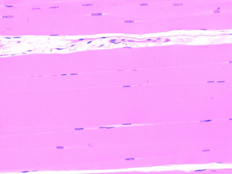
Skeletal muscle is a striated muscle anchored to bones by the piles of collagen fibers described as tendons. Skeletal muscle functions totally under the voluntary regulation of the somatic nervous structure. It stimulates movement, facial utterances, stance, and other volitional locomotions of the body. The muscle cells or myocytes are the fundamental systematic divisions of skeletal muscles. Muscle cells are classified into muscle fibers. Muscle fibers are lengthy, multinucleated, cylindrical cells created from myoblast fusion. Muscle fibers consist of myofibrils made up of thick and delicate myofilaments. The delicate filaments have to do with actin filaments, whereas the thick filaments have to d with myosin filaments. Below the microscope observation, these two filaments are unique banding methods in skeletal muscle. Adding to these two, the muscle fibers also consist of troponin and tropomyosin, which are essential for muscle contraction. The actin and myosin are organized in a recurring division described as a sarcomere. It is the fundamental operational division of muscle fiber and is accountable for the striated presentation. However, the interaction between actin and myosin is accountable for muscle contraction.
What is Cardiac Muscle?
Cardiac muscle is among the three kinds of muscle tissues available in the heart’s walls, mainly in the myocardium of the heart. Comparable to skeletal muscle, the cardiac muscle is as well striated. However, it functions spontaneously, which is unlike the skeletal muscle. The cardiac muscle or the cardiomyocytes are the cells that form the cardiac muscle. These cells possess one, two, or hardly three to four nuclei. The cardiac muscle cells rely on the blood cache to provide oxygen and nutrients and take out the waste product. As a result of the harmonized contraction of heart muscle cells, blood circulation occurs in the circulatory structure. For thorough heart operations, cardiac muscle possesses many mitochondria, several myoglobins, and an efficient blood cache. Cardiac muscles also show cross striations by rotating thick and delicate filaments. Below the electron microscope observations, actin filaments are delicate bands, whereas myosin filaments are thick and darker bands. Cardiac muscle fibers are primarily radiated. T-tubules in the cardiac muscles are more extensive and operate along Z-discs. Intercalated disc links cardiac myocytes to an electrochemical syncitium and are accountable for power communications during muscle contractions. T-tubules handle an essential position in excitation-contraction-coupling known as ECG.
Difference Between Skeletal Muscle and Cardiac Muscle
- Skeletal muscle is described as the striated and evolutionary muscles discovered connected to bones by tendons. Cardiac muscle is described as the striated and spontaneous muscles found in the walls of the vertebrate heart.
- Skeletal muscle is available in every body region, anchored to bones by tendons. Cardiac muscle is available in the heart walls of vertebrates.
- Skeletal muscles stimulate movements, facial utterances, stances, and other voluntary locomotion of the body. Cardiac muscle harmonizes cardiac contractions to discharge blood all through the body and retains blood pressure.
- In skeletal muscles, locomotion is under conscious control. However, this is not so in cardiac muscle.
- Skeletal muscle possesses a mild quantity of ATPase, which is surplus in cardiac muscle.
- Skeletal muscle possesses self-control, but there is no self-control in cardiac muscle.
- Skeletal muscle possesses cylindrical elongated fibers, while cardiac muscle possesses radiated fibers.
- Skeletal muscles consist of several peripheral nuclei, while cardiac muscles consist of just one, two, or three to four centrally sited nuclei.